CALL TODAY for FREE In home consultation
SEND EMAIL
1995 East Norse Avenue
Cudahy, Wisconsin 53110
414-727-2524
4445 W Forest Home Avenue
Milwaukee, Wisconsin 53219
414-727-2525
2140 American Drive
Neenah, Wisconsin 54956
920-257-6060
65 Copeland Avenue
La Crosse, Wisconsin 54603
608-784-9980
Choosing the best skin imaging system for a clinic is not as simple as comparing camera resolution. The right system should support consistent assessments, clear patient communication, and responsible clinical documentation. It must capture details such as pigmentation, redness, texture, pores, and moisture under controlled lighting. Small differences matter.
In daily practice, usability often matters more than impressive specifications. A dermatologist may need fast imaging between appointments, while an aesthetic clinic may value before-and-after comparisons. Look for standardized positioning, repeatable lighting, secure data storage, and software that produces understandable reports. A reliable skin imaging system should fit the clinic’s workflow, not create another complicated task. Staff training is essential. Even advanced equipment can produce misleading results when settings or patient positioning change.
There is no universal winner. Cost, clinical goals, patient volume, maintenance, and available expertise all influence the decision. Independent validation and transparent manufacturer information deserve careful attention. Marketing claims can sound convincing, but they should not replace professional judgment. Some systems may also overemphasize visible changes while missing the broader clinical context. That limitation deserves reflection. The strongest choice combines measurable image quality with ethical use, informed consent, and ongoing review. Clinics should test the system with real cases before committing. A short trial can reveal workflow problems that a product brochure never shows. In the end, the best option is the one that improves observation without pretending to replace experience.
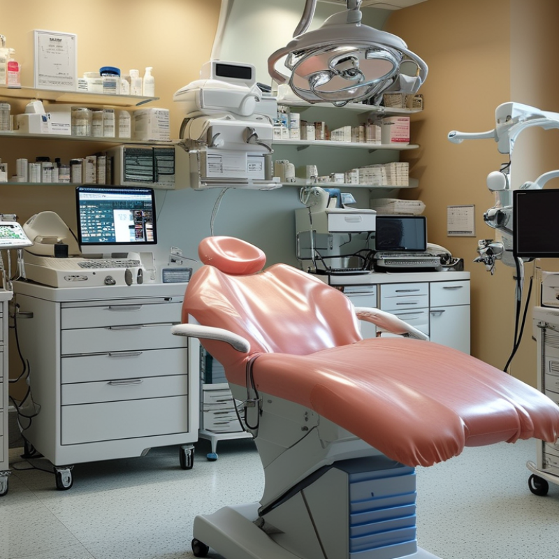
A skin imaging system is a clinical camera setup for examining visible skin changes. It captures standardized photographs under controlled lighting, distance, and angles. This consistency helps clinicians compare pigmentation, redness, texture, pores, and surface irregularities over time. Ordinary phone photos rarely provide the same accuracy.
During an appointment, the practitioner positions the patient’s face or body within a marked area. The patient may rest their chin on a support while several images are taken. Specialized software can then process these images and create detailed maps of tone, texture, oil, or sun-related changes.
Some systems use polarized or ultraviolet-style illumination. These views may reveal patterns that normal lighting hides. Results are more useful when matched with examination findings, medical history, and consistent follow-up images.
Numbers can mislead. Recent makeup, skincare treatments, room humidity, or poor positioning may affect the reading.
A dependable clinic also needs informed consent, secure image storage, regular calibration, and trained staff. Practitioners should check image quality before discussing treatment options. They should explain uncertainty when software produces color maps or risk scores.
The best system is not always the most advanced one. It should suit the clinic’s lighting, workload, training, and follow-up process.
In practice, repeatable images often matter more than impressive screens. No device is flawless. Human judgment remains essential.
The best skin imaging system is not defined by the sharpest picture alone. In clinical practice, consistent images matter more than impressive demonstrations. Look for controlled lighting, fixed camera distance, and clear body-position guides. These details reduce avoidable variation during follow-up visits. A small lighting change can make redness appear worse.
High-resolution images should show surface texture, pigmentation, vessels, and lesion borders. Polarized imaging can reduce glare and reveal deeper visual patterns. Dermoscopic views may support the assessment of suspicious lesions. Three-dimensional imaging can help track volume, contour, and treatment response. However, every feature needs clinical purpose. Extra data can slow consultations without improving decisions.
Reliable software should allow side-by-side comparisons, precise labeling, and secure access controls. Automated analysis may highlight changes, but it should not replace trained clinical judgment. A clinician must review the original image, patient history, and physical findings together. Calibration is easy to overlook. So is staff training. If different employees capture images differently, longitudinal records become less dependable. The system should also export readable reports for multidisciplinary review and patient communication. I would test it during a busy clinic day, not only in a polished product demonstration. No system is perfect. Even strong imaging can mislead when the patient moves, lighting shifts, or the image is interpreted without context.
For routine clinical practice, image standardization, consistent lighting, accurate color reproduction, and reliable side-by-side comparison are usually more valuable than having the largest number of imaging modes. Polarized imaging can reduce surface reflection and improve visualization of subsurface vascular and pigment-related structures, while dermoscopic magnification supports detailed lesion assessment. Three-dimensional measurement is especially useful when monitoring contour, volume, or treatment changes over time.
The scores represent a practical 0–10 clinical-priority framework for selecting an imaging system. They are not manufacturer performance ratings or diagnostic accuracy estimates.
Choosing the best skin imaging system depends on the clinical question, not the most impressive screen. Leading technologies include polarized photography, multispectral imaging, three-dimensional scanning, and high-frequency ultrasound. Each reveals different details.
Polarized images can highlight redness, pores, and surface texture under controlled lighting. Multispectral systems examine pigment and vascular patterns across several wavelengths. They are useful for tracking subtle changes over time.
Three-dimensional scanning measures contours, volume, and asymmetry, which can support procedure planning. Ultrasound explores deeper skin layers, although images may require specialist interpretation.
No system is perfect.
A reliable comparison should examine image repeatability, calibration, software clarity, and staff training. Ask whether the device produces consistent results when a patient returns six months later. Small changes in camera distance or room lighting can distort comparisons. That matters.
Artificial intelligence may flag lesions or patterns, but it should support—not replace—clinical judgment. Independent validation is important, especially when software updates change its performance.
Clinics should also assess secure data storage, patient consent, and export options for medical records. I would not choose a system based only on resolution. Higher detail can expose more noise, and impressive images may still lack diagnostic value.
A practical trial with real patients often reveals workflow problems that a demonstration hides.
The best skin imaging system depends on the clinic’s patients, workflow, and clinical questions. A general dermatology clinic often benefits from polarized and cross-polarized images. These reveal surface texture, redness, pigmentation, and vascular patterns under consistent lighting. A small consultation room may need a compact system with quick capture and simple storage. Speed matters when appointments are tightly scheduled.
Clinics treating acne, scars, or long-term pigmentation need reliable before-and-after documentation. High-resolution photography helps practitioners compare changes across several visits. It also supports clearer patient discussions. Cosmetic clinics may benefit from three-dimensional imaging when assessing facial contour, asymmetry, or treatment planning. Specialist clinics handling suspicious lesions need magnified imaging, stable positioning, and secure clinical records. Image quality alone is not enough. Staff training, calibration, and consistent lighting affect reliability. Small errors matter.
Choose for your workflow.
In practice, expensive equipment can become underused if it takes too long to operate. A simpler system may produce better records because staff use it consistently. However, even consistent images cannot replace clinical examination or professional judgment. Measurements may vary with hydration, lighting, and camera distance. That limitation deserves attention. Before purchasing, clinics should test the system with real patients, review data protection procedures, and assess reporting needs. A useful trial includes busy days, different skin tones, and follow-up visits. One overlooked detail can change the result.
| Imaging System Type | Primary Information Captured | Clinics That Benefit Most | Typical Clinical Uses | Typical Capture Time | Main Strengths | Important Limitations | Best Overall Fit |
|---|---|---|---|---|---|---|---|
| Standardized 2D Photography | Visible-light images of skin surface, color, texture, lesions, redness, and treatment areas. | General dermatology practices, primary-care clinics, aesthetic clinics, and clinics starting a digital imaging program. | Baseline documentation, treatment planning, before-and-after comparison, patient records, and remote consultation support. | Usually less than 1 minute per standardized view, depending on the number of views and workflow. | Low complexity, fast workflow, broad clinical usefulness, and easy image sharing. | Does not directly measure subsurface structures, and results can be affected by lighting, camera angle, distance, and patient positioning. | Best entry-level and general-purpose option |
| Cross-Polarized Photography | Reduced surface glare with improved visualization of erythema, pigmentation differences, and vascular features. | Dermatology, cosmetic dermatology, pigmentation clinics, acne clinics, and practices monitoring redness or inflammation. | Assessment of redness, post-inflammatory pigmentation, uneven tone, acne-related changes, and treatment response. | Usually less than 1 minute per view when integrated into a photographic workflow. | Provides more consistent visualization of color-related features than ordinary room-light photography. | Image appearance depends on calibration and standardized acquisition; it is not a substitute for diagnostic examination or histopathology. | Best for pigment and redness monitoring |
| Ultraviolet or Fluorescence Imaging | Surface fluorescence patterns and selected changes associated with oil, scale, follicular material, or pigment contrast. | Acne-focused clinics, cosmetic practices, skin-care consultation centers, and educational patient-assessment settings. | Patient education, discussion of skin-care routines, assessment of selected surface patterns, and treatment adherence support. | Generally a few minutes, including preparation and image review. | Can reveal contrasts that are less apparent in ordinary visible-light images and can improve patient understanding. | Fluorescence is not specific for one diagnosis; findings can vary with cosmetics, cleansing, skin type, and environmental conditions. | Best as a supplementary educational tool |
| Dermoscopic Imaging | Magnified and illuminated surface structures, including pigment networks, vessels, globules, dots, and lesion architecture. | Dermatology practices, skin-cancer screening clinics, lesion-monitoring services, and clinicians trained in dermoscopy. | Evaluation and documentation of pigmented lesions, inflammatory conditions, hair disorders, and selected nail findings. | Approximately 1–5 minutes per lesion, depending on the number of images and examination complexity. | Shows subsurface-adjacent patterns that are not visible to the unaided eye and supports serial lesion comparison. | Requires clinician training and clinical context; imaging alone cannot confirm malignancy, which may require biopsy and histopathology. | Best for lesion assessment and monitoring |
| 3D Stereophotogrammetry | Three-dimensional facial or body shape, surface contour, volume, symmetry, and selected texture changes. | Plastic-surgery practices, facial-aesthetic clinics, reconstructive services, and high-volume treatment centers. | Pre-treatment planning, volumetric assessment, symmetry analysis, surgical consultation, and objective follow-up. | Usually a few seconds for image capture, with additional time for positioning, processing, and analysis. | Supports quantitative comparison of contour, volume, and symmetry rather than relying only on visual judgment. | Higher purchase and training requirements; hair, movement, reflective surfaces, and inconsistent positioning can reduce measurement reliability. | Best for procedures involving shape or volume |
| High-Frequency Ultrasound | Ultrasound echoes from superficial tissue layers, allowing assessment of skin thickness and selected subcutaneous structures. | Specialist dermatology, scar-management services, research-oriented clinics, and practices evaluating superficial tissue changes. | Assessment of selected scars, edema, skin thickness, superficial masses, and treatment-related tissue changes. | Often 5–15 minutes for a focused examination, depending on the anatomical site and protocol. | Provides depth-related information that photographic systems cannot provide and does not use ionizing radiation. | Requires trained operators, suitable contact technique, and interpretation within the clinical context; superficial resolution varies by frequency. | Best for superficial tissue-depth assessment |
| Optical Coherence Tomography | High-resolution cross-sectional optical images of superficial skin microstructure. | Academic medical centers, specialist dermatology units, clinical research programs, and clinics with advanced diagnostic workflows. | Non-invasive structural assessment, research imaging, monitoring of selected lesions, and evaluation of superficial treatment effects. | Commonly several minutes per target area, depending on scanning protocol and interpretation needs. | Offers cross-sectional structural information without biopsy or ionizing radiation. | Limited penetration depth, higher cost, specialized interpretation, and availability constraints can restrict routine use. | Best for advanced diagnostics and research |
| Multimodal Imaging Platform | A combination of visible-light, polarized, ultraviolet, dermoscopic, or three-dimensional image data in one documented workflow. | Large dermatology groups, multidisciplinary practices, aesthetic centers, clinical trial sites, and clinics with high documentation requirements. | Comprehensive baseline records, lesion monitoring, cosmetic treatment planning, outcome measurement, and longitudinal patient management. | Typically several minutes per patient, depending on the selected imaging modes and number of anatomical sites. | Combines complementary information and can improve consistency when positioning, lighting, and documentation are standardized. | Higher acquisition cost, staff-training needs, data-storage requirements, and workflow complexity. | Best for high-volume and comprehensive clinics |
Selecting the best skin imaging system starts with clinical purpose, not the sharpest promotional image. A dermatology clinic may need lesion tracking, while an aesthetic practice may prioritize standardized before-and-after photography. Define the conditions, body sites, and review intervals your team actually handles. Ask for repeatability. During a demonstration, photograph the same volunteer under identical distance, lighting, and positioning. Compare color balance, focus, measurement tools, and image retrieval speed. A vivid image is not automatically a useful image. That distinction matters.
Implementation should begin with a controlled pilot, not a clinic-wide purchase. Include clinicians, nurses, photographers, and administrative staff in the evaluation. They will reveal practical problems that specifications often hide. Check whether the system supports calibrated lighting, consistent patient positioning, secure access, and clear audit records. Confirm how images integrate with existing records and whether data can be exported in a usable format. Patient consent, privacy controls, retention periods, and local regulatory requirements need written procedures. Training should include image capture, quality checks, troubleshooting, and appropriate interpretation. Never treat an image as a diagnosis by itself.
A reliable clinic also measures performance after installation. Track repeat visits, rejected images, staff time, and patient communication errors. Review these results after several weeks. The first workflow may feel inefficient. That is normal. It may also expose weak assumptions about room layout or staff availability. Keep a small library of quality examples, including imperfect images, for training. Schedule calibration checks and software reviews. Ask an independent clinical or technical adviser to examine the protocol when possible. The best system is the one your team can use consistently, explain clearly, and maintain responsibly.
1995 E Norse Ave
Cudahy, WI 53110
Phone: 414-486-6249
Fax: 414-486-5728